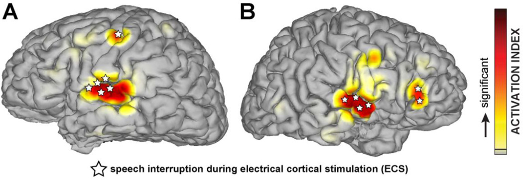
Fig. 2.
Cortical representation of receptive language in left (A) and right (B) hemispheres identified through the electrocorticographic (70–170 Hz broadband gamma) response to a passive listening task (red shading) and speech interruption during ECS (white stars). The results from both methods are congruent and identify language-related areas in central superior temporal gyrus, Broca’s area, and pre-motor cortex.

