
Figure 5.
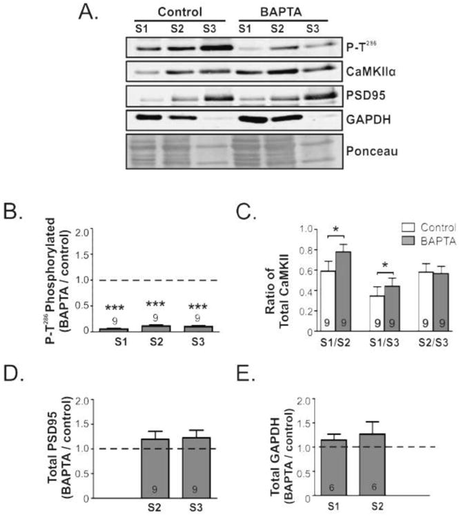
Depletion of extracellular calcium induces CaMKII translocation to the cytosolic fraction. Mouse brain slices were incubated for 15 min with BAPTA (5 mM) or vehicle. Equal volumes of cytosolic (S1), membrane (S2, Triton-Soluble), and synaptic (S3, Triton-Insoluble) fractions isolated from dorsal striatum were immunoblotted for: P-Thr286, total CaMKIIα, and also GAPDH and PSD-95 as cytosolic and synaptic markers, respectively. Representative images of each blot are shown, as well as a segment of the Ponceau stained membrane prior to immunoblotting. The relative enrichment of the P-Thr286/total CaMKIIα ratio (B) as well as the total CaMKIIα (C) PSD95 (D) GAPDH (E) normalized to the Ponceau stain in each fraction from control and BAPTA treated slices were then quantified. All bar graphs report the mean ± SEM from the indicated number of replicates. The dashed line indicates a value of 1.0 (F); the theoretical value if BAPTA had no effect. Significance determined by one sample -test (*p<0.05, ***p<0.001).