
Fig. 2.
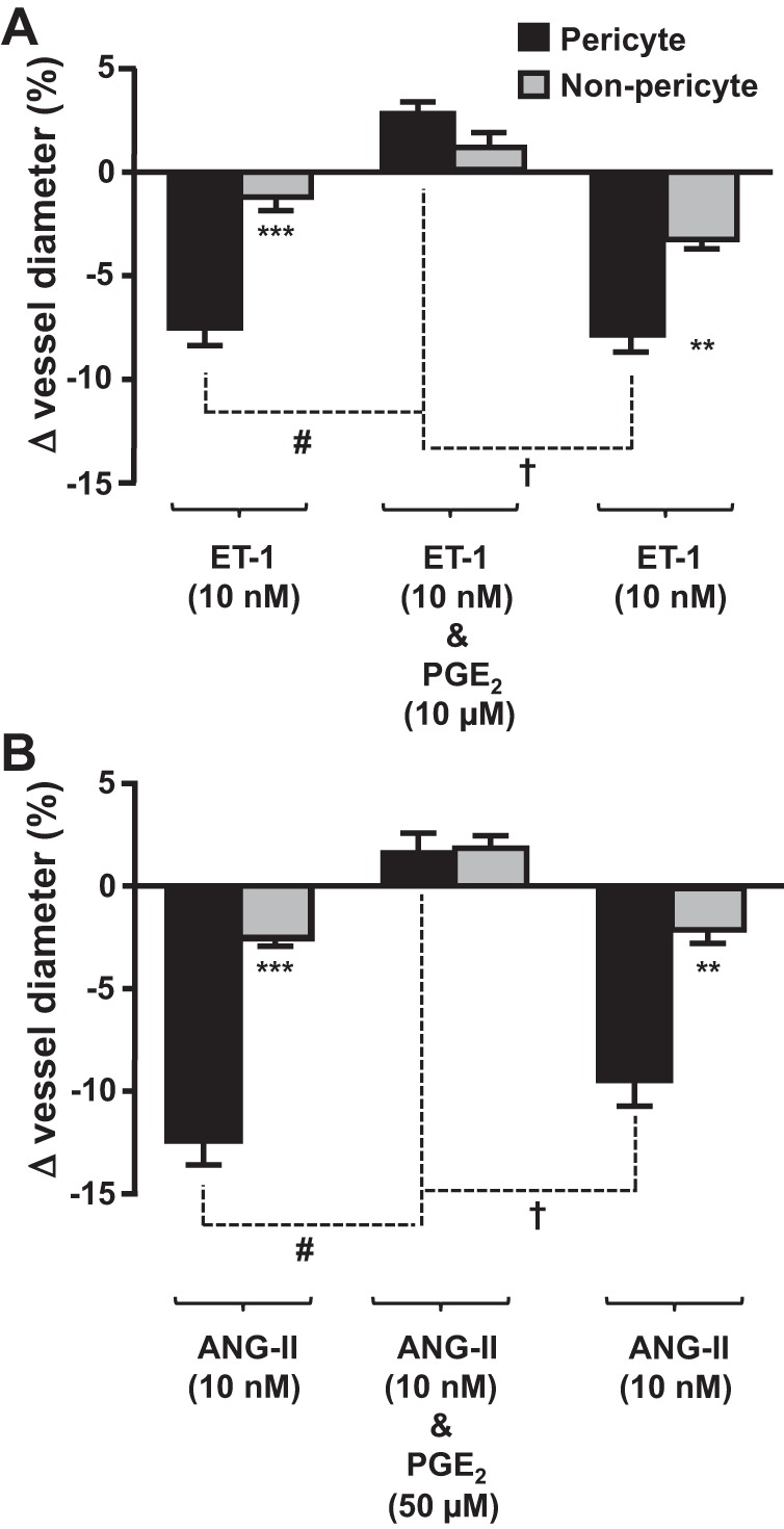
PGE2 attenuates the endothelin (ET)-1- and ANG II-evoked vasoconstriction of in situ vasa recta by pericytes. Bar graphs show percent changes in vasa recta diameter at pericyte and nonpericyte sites. ET-1 (10 nM; A) and ANG II (10 nM; B) evoked significantly (***) greater constriction at pericytes sites than at nonpericyte sites. The addition of PGE2 [10 μM (A) and 50 μM (B)] significantly (#) attenuated the pericyte-mediated vasoconstriction evoked by ET-1and ANG II, respectively. After removal of PGE2, ET-1 and ANG II alone evoked a second vasoconstriction, indicating that the effect of PGE2 was reversible. No significant change in vessel diameter was observed at nonpericyte sites. Values are means ± SE; n ≥ 6 slices and n ≥ 4 animals. **P < 0.01; ***P < 0.001; #P < 0.05; †P < 0.05.