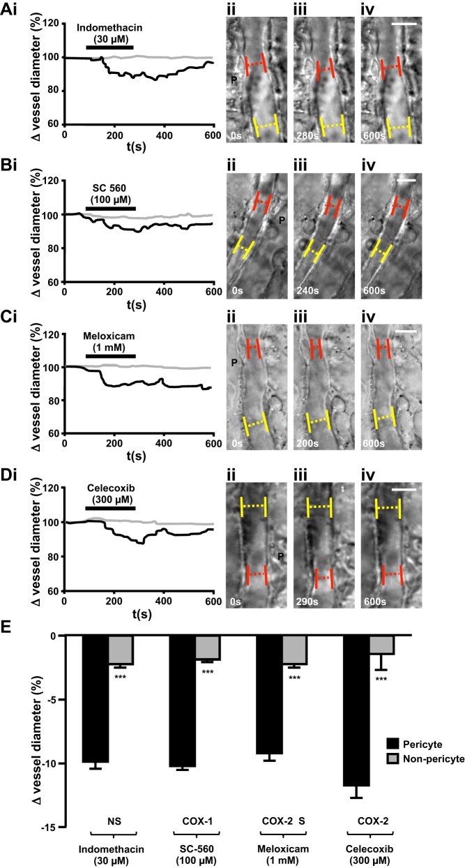
Fig. 3.
Superfusion of live kidney tissue with nonselective nonsteroidal anti-inflammatory drugs (NSAIDs) caused pericyte-mediated constriction of vasa recta. Representative traces (A,i–D,i) of percent changes in vessel diameter recorded over time at pericyte sites (black lines) and nonpericyte sites (shaded lines) when kidney slices were exposed to either indomethacin (30 μM; A,i), SC-560 (100 μM; B,i), meloxicam (1 mM; C,i), or celecoxib (300 μM; D,i) are shown. Corresponding differential interference contrast images of pericyte (P) and nonpericyte sites before drug exposure (ii), during superfusion of the drug (iii), and after exposure, during washout of the drug (iv), for each NSAID used [indomethacin (A), SC-560 (B), meloxicam (C), and celecoxib (D)] are also shown. Pericytes are denoted by black dotted circles; red dotted lines and yellow dotted lines indicate where changes in vessel diameter were measured at pericyte sites and nonpericyte sites, respectively. E: superfusion of either indomethacin (30 μM), SC-560 (100 μM), meloxicam (1 mM), or celecoxib (300 μM) onto live kidney slices evoked a significantly greater constriction of vasa recta at pericyte sites compared with nonpericyte sites. NS, nonselective; COX-1, cyclooxygenase-1 specific; COX-2 S, cyclooxygenase-2 selective; COX-2, cyclooxygenase-2 specific. Values are means ± SE; n ≥ 5 slices and n ≥ 4 animals. ***P < 0.001.

