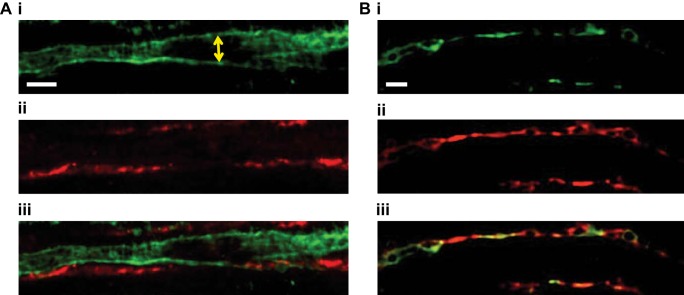
Fig. 9.
Confocal images of EP2 and EP4 receptor expression in the renal medulla. Before fixation with paraformaldehyde, vasa recta capillaries of live kidney slices were labeled with Alexa 488-conjugated IB4 antibody (green; A,i and B,i). A,ii: EP2 receptor expression was labeled with anti-EP2 primary antibody and probed with Alexa 555-conjugated secondary antibody (red). A,iii: overlay of images of A,i and ii, showing the close proximity of vasa recta (green) and EP2 receptor expression (red). B,ii: EP4 receptor expression was labeled with anti-EP4 primary antibody and probed with Alexa 555-conjugated secondary antibody (red). B,iii: overlay of images of B,i and ii, suggesting colocalization of vasa recta (green) and EP4 receptor expression (red). n = 6 slices and n = 3 animals.

