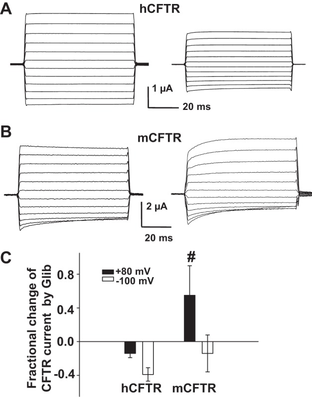
Fig. 11.

Effects of glibenclamide on hCFTR and mCFTR currents in whole oocytes. Families of TEVC currents in the absence (control, left) and presence (right) of 100 μM extracellular glibenclamide for hCFTR (A) and mCFTR (B). The voltage protocol included a holding potential VM = −30 mV, then membrane potential stepped to potentials ranging from VM = −140 mV to +80 mV in 20-mV increments with a period of 75 ms at each voltage. C: summary of the fractional change in hCFTR and mCFTR current at VM = +80 mV and −100 mV. #P < 0.01 compared with hCFTR at VM = +80 mV; n = 4–5 for each condition.
