Figure 3.
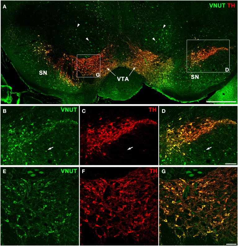
VNUT-immunoreactive cells co-localize with tyrosine-hydroxylase positive dopaminergic neurons in the mouse substantia nigra and ventral tegmental area. (A) A coronal section of the mouse midbrain co-labeled with VNUT (green) and Tyrosine Hydroxylase (TH, red) showing co-localization on dopaminergic neurons in the substantia nigra (SN) and ventral tegmental area (VTA). Arrowheads indicate VNUT+TH− cells. (B–D) A magnified view of the SN (right inset) showing all TH-positive cells (red) are also positive for VNUT (green). Arrows in (B–D) indicate VNUT+TH− cells. (E–G) A magnified view of the VTA (left inset) showing VNUT expression (green) in all TH-positive cells (red). VNUT expression was not restricted to DA neurons and was found in many TH-negative cells throughout this region of the brain. Scale bars = 500 μm (A), 100 μm (B–D) and 50 μm (E–G).

