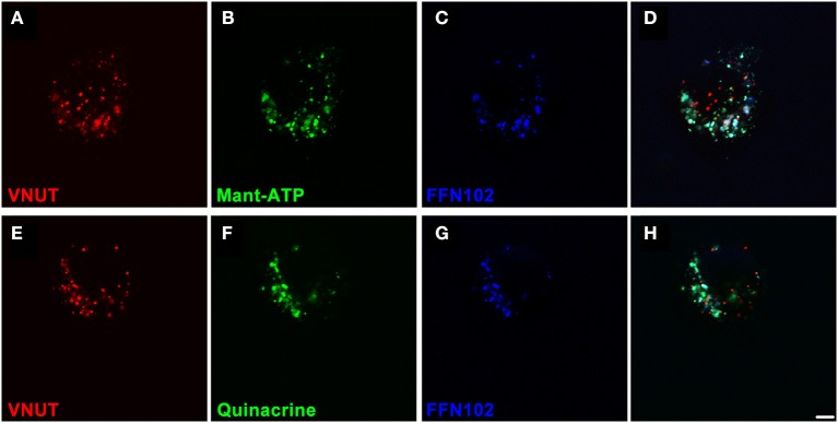
Figure 5.

VNUT-immunoreactive dissociated retinal neurons co-load fluorescent ATP- and dopamine-markers in vesicles. Freshly dissociated retinal preparations were labeled for VNUT-immunoreactivity (red) and incubated with fluorescent ATP markers (green) and the fluorescent dopamine marker (FFN102, blue). (A–D) VNUT-positive neurons (A, red) co-expressed the fluorescent ATP marker Mant-ATP (B, green) and the dopamine fluorophore FFN102 (C, blue) in discrete puncta (D, co-localized). (E–H) Similarly VNUT-positive neurons (E, red) co-expressed the fluorescent ATP marker quinacrine (F, green) and the dopamine fluorophore FFN102 (G, blue) in discrete puncta (H, co-localized). Within these cells, the fluorescent ATP marker puncta co-localized with the dopamine fluorophore, FFN102, suggesting co-loading of ATP and dopamine in vesicles within VNUT-positive neurons. Scale bar = 2.5 μm.
