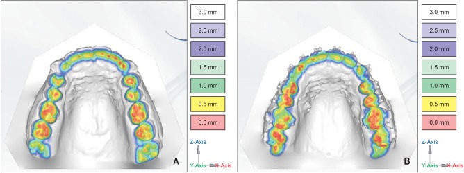
Figure 3. Evaluation methods of vertical teeth movement by three-dimensional digital model. A, Before treatment; B, after treatment. The red areas are contact areas, which are more marked on the right first maxillary molars before treatment than after treatment. Red marks are stronger on premolars and second molars after treatment, which is an additional sign of first molar intrusion.

