
FIG. 1.
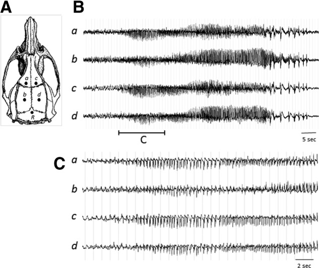
(A) Illustration of EEG electrode configuration. Electrodes a–d were individually referenced to the electrode R to create a four-channel monoreferential EEG recording montage. (B) An example of NCS episode detected by EEG. (C) Seizure initiation shown on an expanded timescale. EEG, electroencephalographic; NCS, nonconvulsive seizures.