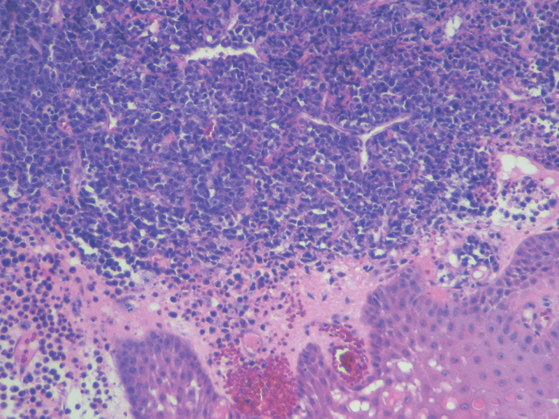
Fig. 5.

Oral mucosal lesion biopsy revealed the existence of an atypical and dense infiltrate, relativity uniform, composed of large cells with a moderate cytoplasm, an eccentric nuclei, and one or more large nucleoli. Also, it is possible to see a variable number of small cells with plasmacytic appearance, consistent with the diagnosis of high-grade non-Hodgkin lymphoma. Hematoxylin and eosin, ×200.
