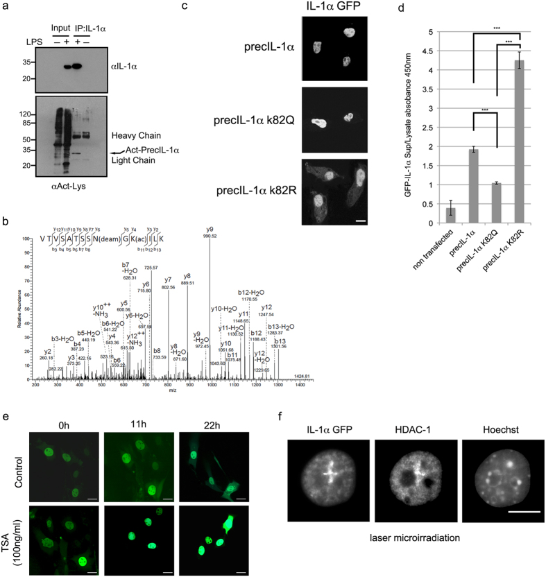
Figure 3. IL-1α acetylation within the nuclear localization sequence impacts on IL1α subcellular localisation.
(a) IL-1α precursor is recognized by a pan acetyl antibody. Endogenous IL-1α was immunoprecipitated (IP) from nuclear extracts of Raw 264.7 cells, either induced or non-induced with 100 ng/ml LPS. Total IP proteins were separated over 15% SDS PAGE, transferred to nitrocellulose membranes and blotted with anti-mouse IL-1α (top panel) or anti-Kac (bottom panel). Acetylated IL-1α is marked by arrows and IP antibody light and heavy chain signals are indicated. (b) Annotated MS/MS spectrum of the tryptic peptide VTVSATSSN(Deam)GK(Acetyl)ILK (MH2 + 724.40 Da) showing acetylation of IL-1α (Uniprot ID P01582) at K82 and N80 deamidation. (c) PrecIL-1α K82 mutants affect IL-1α sub-cellular localization. Confocal microscopic analysis of GFP tagged WT IL-1α and mutations of precIL-1α K82 to glutamine (precIL-1α K82Q, mimicking acetylation) and to arginine (precIL-1α K82R non-acetylateable). White scale bars, 20 μm (d) IL-1α K82 mutations reduce cytokine secretion after DNA damage. Mouse B16 cells were transfected with the indicated GFP IL-1α plasmids. The cells were then subjected to 100 μM H2O2. 16h after stress induction levels of secreted GFP IL-1α in cell growth medium was measured using a GFP ELISA. GFP IL-1α levels in cell lysates were used to normalize for transfection efficiencies and non-transfected cells were used as negative controls. Data are expressed as mean ± SD of three independent experiments. (e) Histone deacetylase inhibition by TSA increases IL-1α nuclear localization. Images of cells expressing GFP IL-1α either non-treated (control) or treated with TSA (100 ng/ml) were collected every hour for 22 h and representative images for three time points (0, 11 and 22 hours) are shown (For averaged fluorescence intensities of nuclear/cytoplasmic ratios see Supplementary Figure 1b). (f) HDAC-1 and IL-1α can co-localize at DNA damage lesions. Cells expressing GFP IL-1α were laser-microirradiated for the induction of DNA damage. Localization of HDAC-1 and IL-1α–GFP were visualized by confocal microscopic analysis.

