FIGURE 1.

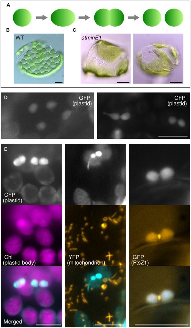
Utility of cyan fluorescent protein (CFP) to investigate plastid morphology in Arabidopsis leaf epidermis. (A–C) A framework describing the replication and morphology of leaf mesophyll chloroplasts. Schematic diagram of chloroplast replication by binary fission (A) and chloroplast phenotypes in WT (B) and atminE1 (C) leaf mesophyll cells are shown. (D) Detection of plastid-targeted green fluorescent protein (GFP, left) and CFP (right) in leaf epidermis. (E) Dual detection of plastid-targeted CFP and chlorophyll (Chl magenta-colored), mitochondria-targeted YFP (orange-colored) or FtsZ1–GFP (orange-colored) in leaf epidermis. (D,E) Leaves from 2-week-old seedlings of WT-background transgenic lines were observed by fluorescence microscopy. In merged images, CFP fluorescence is colored in cyan. Bars = 5 μm (black) and 10 μm (white).
