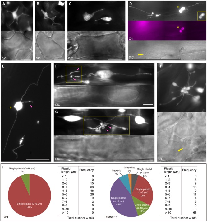
FIGURE 3.

Various types of plastid morphology and distribution patterns in leaf epidermis of atminE1. (A–H) Images of CFP-labeled plastids in leaf petiole epidermis of 2- or 3-week-old atminE1 seedlings. DIC and chlorophyll fluorescence (Chl magenta-colored) images are also shown. Asterisks indicate plastids with chlorophyll signals only within cells, while double arrowheads indicate cell nuclei. Single arrowheads and boxes represent plastid bulges associated with plastid bodies or stromules and their activated regions, respectively. Inset in (D) is a CFP image of a chlorophyll-positive plastid pair taken using a shorter exposure time. Bars = 10 μm (white) and 100 μm (black). (I) Measurement of plastid morphologies in WT and atminE1. Phenotypes of epidermal plastids in 2-week-old seedlings were classified into three groups, ‘single plastid’, ‘network’ and ‘grape-like’. The former group was further examined with respect to total plastid length. Plastid length was defined as the length of the longest line passing over the plastid area. Since the borders between plastid bodies and stromules were often unclear in atminE1, the plastid length includes the area of stromules (for both WT and atminE1 samples).
