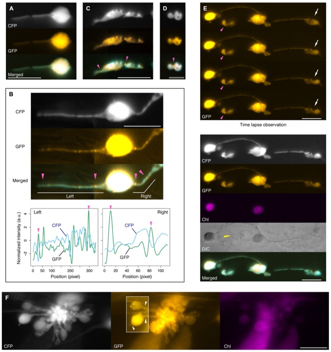
FIGURE 6.

FtsZ1 localization in stromules and plastid bulges in leaf epidermis of atminE1. (A–F) Dual detection of FtsZ1–GFP and stroma-targeted CFP in epidermal plastids of atminE1 leaf petioles. Fluorescence images of CFP, GFP (orange-colored), chlorophyll (magenta-colored), and merged (CFP cyan-colored, GFP orange-colored) are shown. In (B), line profiles of normalized CFP and GFP signal intensity in left and right stromules in the image are also presented. Single arrowheads indicate position of FtsZ1 ring placement, while box in (F) highlights FtsZ1 ring placement at stromules or between plastid body and plastid vesicle within a grape-like plastid association. Arrows and a double arrowhead in (E) indicate a plastid constriction event by time-lapse microscopy and the position of cell nucleus, respectively. Bars = 5 μm (D), and 10 μm (others).
