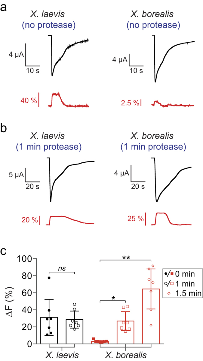
Figure 5. Effect of protease treatment on fluorescence signals (ΔF) from X. laevis and X. borealis oocytes expressing the α1N203C GlyR with current induced by 10 μM glycine.

(a) Representative current and fluorescence traces show a significantly greater ΔF in untreated X. laevis than X. borealis oocytes. (b) A significant increase in ΔF was obtained after 1 min treatment with protease for X. borealis, but not X. laevis, oocytes. Traces from A and B represent separate oocytes. (c) ΔF of X. laevis and X. borealis oocytes in all conditions tested (n = 7). X. laevis oocytes were not amenable to protease treatment for >1 min as this damaged membrane integrity. P values were calculated in comparison to untreated oocytes from each species using an unpaired t-test with Welch’s correction (*P < 0.01, **P < 0.001). Error bars indicate 95% confidence intervals.
