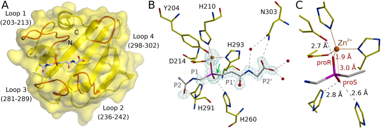
Figure 1. The binding mode of the phosphinate.
(A) The overall structure of the LytM catalytic domain in complex with the transition state analogue. The loops lining the LytM substrate binding cleft are indicated in red. (B) The interactions of the transition state analogue with the protein. The map is the composite omit map contoured at 2.0 Å rmsd. (C) Detail of the phosphinate binding mode (only the P1 and P1′residues excluding the carbonyl are shown). Note that the proR and proS assignment is according to IUPAC priorities for the transition state and not for the phosphinate itself.

