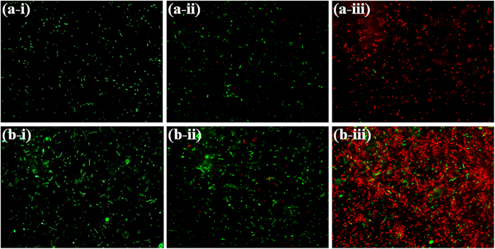
Figure 6. Fluorescence microscopic images of B. subtilis and E. coli in absence and presence of n-IONP and p-IONP.
Intact B. subtilis (a-i), B. subtilis in presence of 50 μM of n-IONP (a-ii), and B. subtilis in presence of 50 μM of p-IONP (a-iii), intact E. coli (b-i), E. coli in presence of 50 μM of n-IONP (b-ii), and E. coli in presence of 50 μM of p-IONP (b-iii). The scale bars represent for 20 μm.

