. 2015 Oct 5;44(20):7342–7370. doi: 10.1039/c5cs00376h

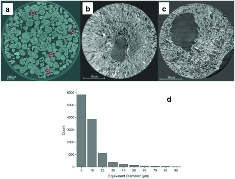
Fig. 24. (a) Single slice of the X-ray micro-tomogram of E-cat particles inside a polyimide tube, which is the circle around the image. The E-cat catalyst particles have a range of shapes and sizes, and some are hollow. Some of them are indicated by the red circles in the image; (b and c) 2D transmission X-ray microscopy images of sections of two E-cat particles showing different sizes of internal voids, which are the dark colored regions in the images; and (d) equivalent diameter of internal voids in the set of E-cat catalyst particles as investigated with X-ray micro-tomography. (Reproduced with permission from ref. 172, Copyright Wiley-VCH, 2014).

Fig. 24