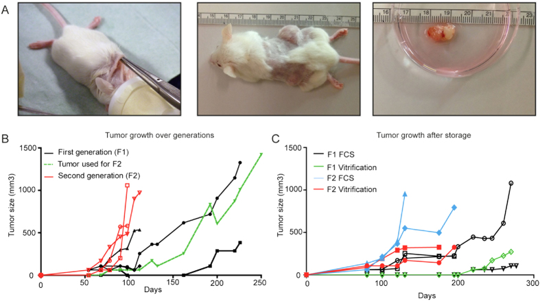
Figure 1. Establishment of the ovarian cancer PDX model.
(A) Making a single cut in the neck, two pieces were subcutaneously transferred to and implanted on either side of the flank of 6–12 weeks old female NOD.Cg-Prkdcscid Il2rgtm1Wjl/SzJ mice. Tumours were measured once or twice a week and after reaching appropriate size, tumours were harvested for either direct propagation into a further generation or for storage. (B) Tumour growth of fresh implanted tumour tissue from patient 56 and further propagation of the tumour (green line) into the second generation (red lines). (C) Tumour growth of stored and subsequently thawed and re-implanted tumour tissue from patient 56. Tumour tissue was either directly frozen after patients primary surgery (F1) using either the vitrification (green line) or FCS/DMSO (black line) protocol. After establishment of a PDX, tumour tissue was harvested from the mouse (F2) and frozen using either the vitrification (red line) or FCS/DMSO (blue line) protocol.

