
Figure 3. Immunohistochemistry of the ER and PR in representative slides of the primary tumour tissue of patient 56 (Patient), tumour tissue from F2 generation of PDX 56 (F2), and tumour tissue from F2 generation of cryopreserved PDX 56 F1 using either FCS/DMSO (F2:FCS) or vitrification (F2:Vitri).
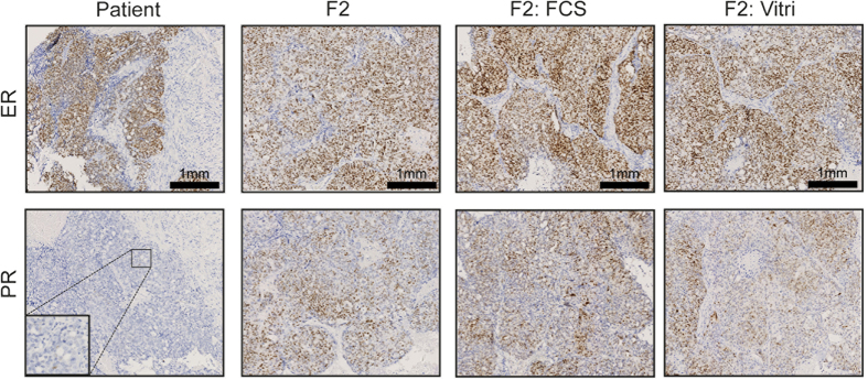
Magnification 10× and 40×.