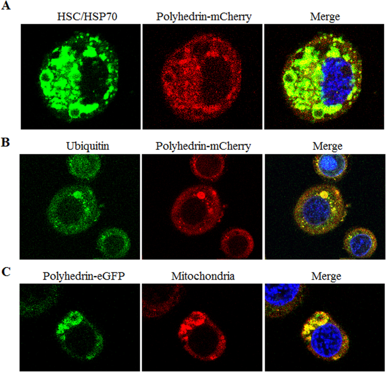
Figure 5. Colocalization analyses of aggregated polyhedrin with HSC/HSP70s, ubiquitinated proteins and mitochondria.
Infected cells expressing polyhedrin-mCherry and polyhedrin-eGFP were collected at 24 h p.i., treated with paraformaldehyde and Triton X-100, and then immunostained for HSC/HSP70s (A) and ubiquitin (B). The mitochondria (C) were stained with a MitoTracker® deep red probe (Molecular Probes, Life technologies, Eugene, OR, USA). Cell nuclei were stained with Hoechst 33342. Cells were imaged under a Leica TCS SP5 confocal laser-scanning microscope.

