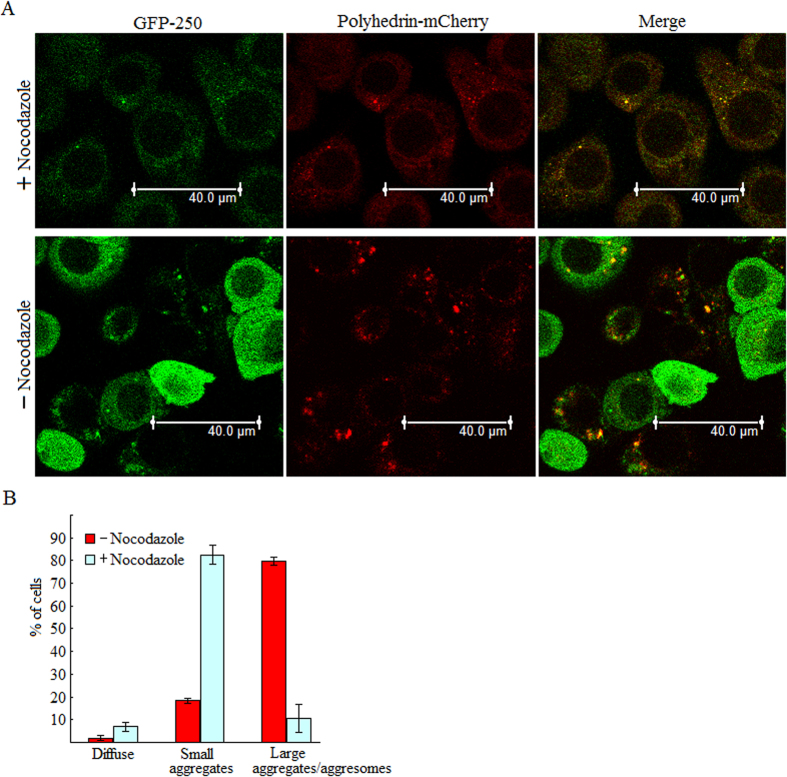
Figure 6. Formation of polyhedrin foci is microtubule-dependent.
(A) Microtubule disruption prevents the formation of coalesced polyhedrin aggregates. BmN cells were co-infected with viral stocks individually expressing GFP-250 and polyhedrin-mCherry, treated with nocodazole at 0 h p.i., and then imaged by a confocal fluorescence microscopy at 24 h p.i. (B) Bar graph describing the effect of nocodazole on polyhedrin aggregate size. Each value is summarized from three independent infections. For each infection, 90–143 cells were scored. Error bars indicates ±SEM.

