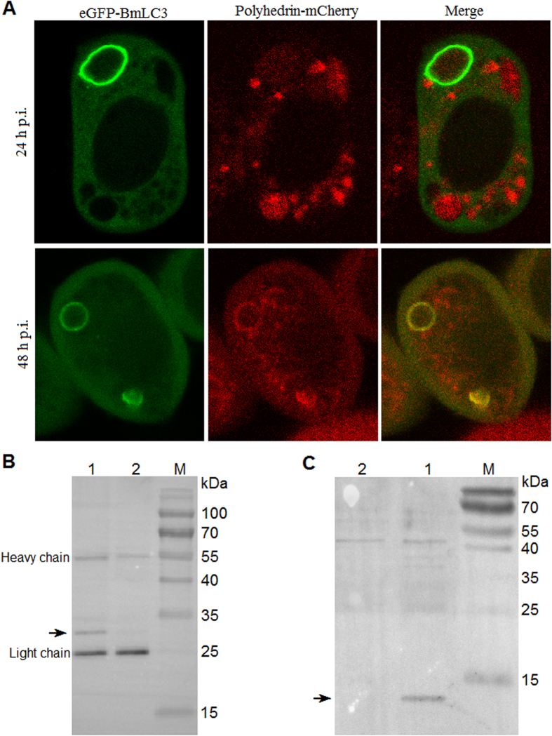
Figure 7. Polyhedrin interacts and is colocalized with BmLC3 on the isolation membrane of autophagosome.
(A) Subcellular localizations of polyhedrin-mCherry and eGFP-BmLC3. The spherical structures indicated autophagosomes. Polyhedrin-mCherry was not at 24 h p.i. (upper panel) but at 48 h p.i. (lower panel) colocalized with BmLC3 on the isolation membrane of autophagosome. (B) eGFP-BmLC3 co-immunoprecipitates with polyhedrin. Plasmids pPie1-eGFP-BmLC3 and pPie1-eGFP were applied for transposition and transfection. BmN cells were co-infected with equal MOI (10 TCID50/cell) of a P2 viral stock and the BmNPV T3 isolate. eGFP or eGFP fused BmLC3 were immunoprecipitated from total cellular extract with a μMACS GFP Isolation Kit and subjected to SDS-PAGE. Co-purified polyhedrin was detected by immunoblotting with a mouse monoclonal anti-polyhedrin antibody. Lane M, protein marker; lane 1, immunoprecipitate from cells co-infected with BmNPV T3 isolate and the viral stock expressing eGFP-BmLC3; lane 2, immunoprecipitate from cells co-infected with BmNPV T3 isolate and the stock expressing eGFP. Arrow indicated the polyhedrin. (C) Polyhedrin-eGFP co-immunoprecipitates with BmLC3. Vectors pPph-Polyhedrin-eGFP and pPph-eGFP were used to obtain individual viral stock. Polyhedrin-eGFP and eGFP alone were co-immunoprecipitated for SDS-PAGE and then Western blot with a rabbit polyclonal anti-BmLC3 antibody. Lane M, protein marker; lane 1, immunoprecipitate from cells infected with the viral stock expressing polyhedrin-eGFP; lane 2, immunoprecipitate from cells infected with the virus expressing eGFP. Arrow indicated BmLC3.

