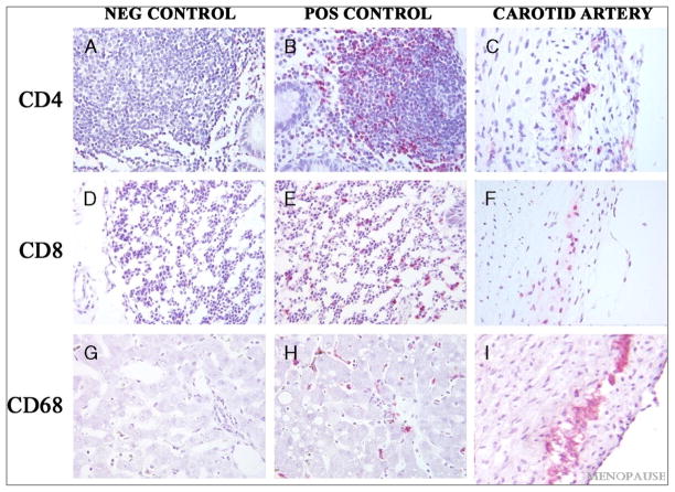
FIG. 1.
Immunohistochemical staining of CD4, CD8, and CD68 in sections of monkey carotid artery along with positive and negative controls: Negative Controls (A, D, G); Positive Controls (B, E, H); Arterial tissues (C, F, I). A–C, Immunostaining for CD-4; D–F, Immunostaininig for CD8; G–I, Immunostaining for CD68. Mucosa associated lymphoid tissue from cynomolgus macaque colon was used as the control tissue for CD4 and CD8, liver was used as the control tissue for CD68. Sections depicted in the first column (A, D, G) were incubated with normal mouse serum instead of primary antibody. All figures at 40X magnification.

