Figure 5.
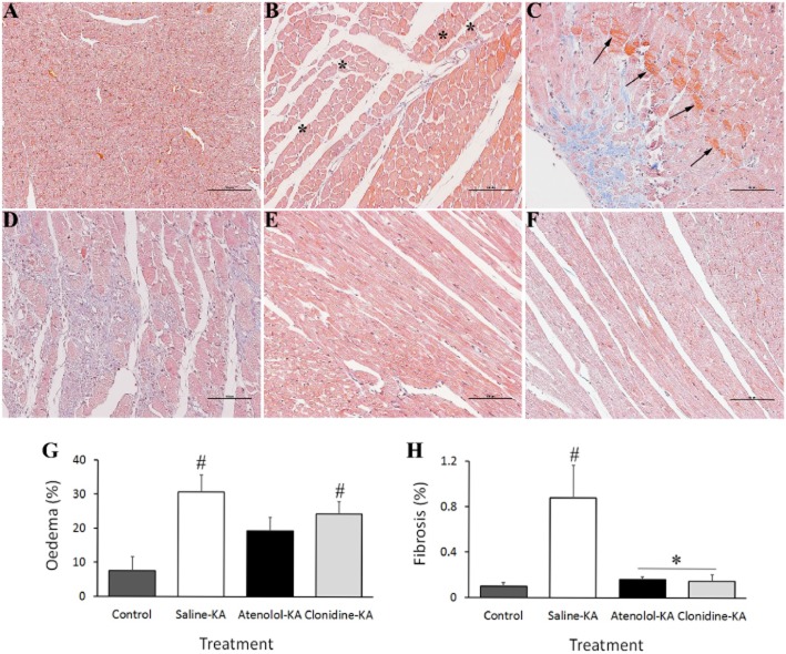
Representative micrographs of left ventricular subendocardium collected at 48 h following (A) saline administration to control animals showing normal representative myocardium; (B–F) Effects of KA administration showing: (B) myocyte vacuolization (asterisks indicate intracellular vacuoles displacing nuclei) and oedema; (C) Coagulative myocytolysis illustrated by hypercontraction band necrosis associated with fibre derangement (arrowheads) and fibrosis; (D) Inflammatory necrosis encapsulated by collagen deposition (blue fibres) indicative of early restorative fibrosis. Hearts from atenolol (E) and clonidine (F) pretreated animals showing preserved normal cardiac morphology. The percentage of tissue positively stained for oedema (G) and fibrosis (H) recorded in 10 sections taken 6 mm from the apex. *P < 0.05 compared with Control-Saline, #P < 0.05 compared with Saline-KA; one-way anova with Dunnett test

