Figure 2.

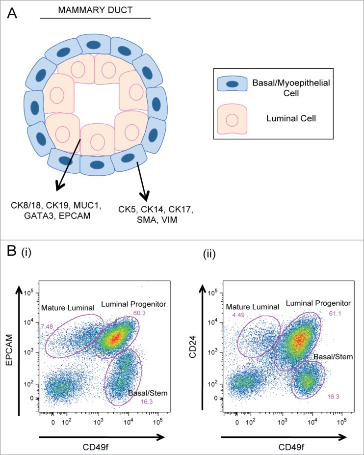
Cellular components of the mammary epithelium. (A) Cross section of a bilayered duct. Luminal cells line the inner duct lumen and are surrounded by an outer layer of contractile basal cells. (B) Flow cytometry plots of primary human mammary epithelial cells analyzed for expression of (i) EPCAM vs. CD49f and (ii) CD24 vs. CD49f expression. Mature luminal, luminal progenitor, and basal/stem populations are indicated.
