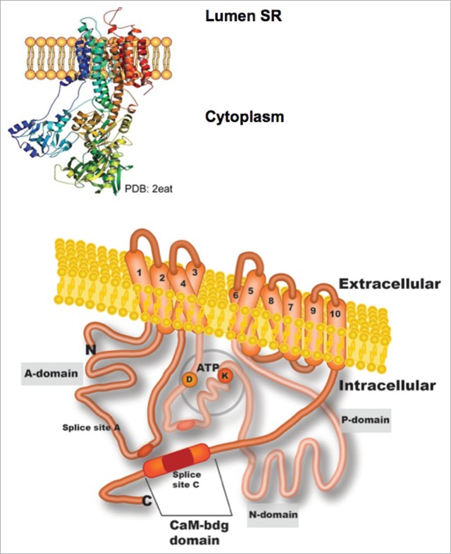
Figure 1.
Illustrated topology of a plasma membrane Ca2+-ATPase (PMCA). The three-dimensional representation of reticulum sarcoplasmic Ca2+-ATPase (SERCA; PDB:2eat) that is shown at the top of the figure was used to construct the PMCA model below. The structure predicted for PMCA includes most of the domains being oriented toward the cytoplasmic face and 10 transmembrane domains (TM). Domain A, which is between TM domains 2 and 3, contains splice site A and a site for phospholipid binding. The intracellular loop located between TM domains 4 and 5 comprises the P- and N-domains where phosphorylation of an aspartate residue (D) and ATP-binding of a lysine residue (K) occur. The C-terminus also contains a calmodulin-binding site in splice site C.

