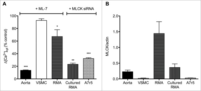
Figure 1.

Effect of MLCK inhibition on agonist-induced cytosolic Ca2+ concentration ([Ca2+]cyt) increase in comparison with MLCK expression level in aorta, cultured aortic smooth muscle cells (VSMC), resistance mesenteric artery (RMA), cultured RMA (organ-cultured for 4 days) and the aortic cell line A7r5. (A) Δ[Ca2+]cyt response after agonist stimulation (noradrenaline 10 μM in RMA and cultured RMA and vasopressin 10 nM in aorta, VSMC and A7r5 cells) in the presence of the MLCK inhibitor (ML-7 at 3 μM in aorta and RMA and 10 μM in VSMC) or after MLCK down-regulation with anti-MLCK-siRNA (in cultured RMA and A7r5) compared to control stimulated conditions. The VOC channel antagonist verapamil (1 μM) was used to block the voltage-dependent component of the [Ca2+]cyt increase in response to vasopressin in aorta. (B) Mean values of Western blot data. MLCK expression was normalized to the actin content. Vertical bars represent the SEM (n = 3–7). *, **, *** Significant differences compared to control conditions. Results were extracted from published data.10,47 The expression level of MLCK is lower in VSMC compared to whole vascular smooth muscle (aorta or RMA) but this does not explain the weaker contribution of MLCK to Ca2+ channel regulation in VSMC. Indeed larger inhibition of Δ[Ca2+]cyt is observed in aorta compared to RMA, while the expression level of MLCK is lower.
