Figure 2.
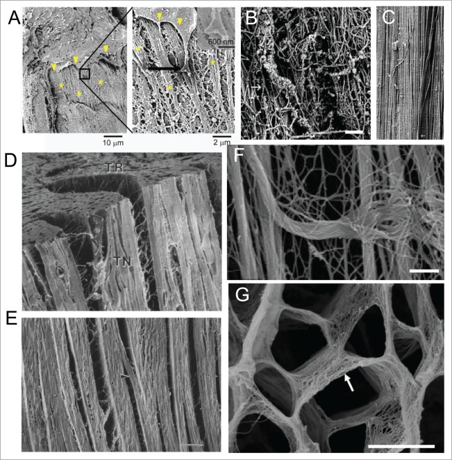
Nanotopographical features are diverse in their shapes and sizes. (A) SEM image showing interaction of an endothelial cell in the aorta with the basement membrane61; left image shows the edge of the cell (arrowhead) in close proximity with the rough matrix in the basement membrane (denoted by *), boxed area magnified in right shows end process of the cell interacting with the basement membrane composed of ridges, bumps, pits and other features in nano- to micro scales; Reprinted with the permission of Mary Ann Liebert, Inc. (B–C) Topographical features dynamically change during development to assume their mature shape in an adult tissue16; (B) SEM image of late embryonic bovine medial collateral tendon ligament show substantial interwoven collagen fibrils, arranging themselves toward the long axis; (C) Mature tendon ligament show a highly aligned 3D network of collagen fibrils along the long axis of the ligament, with only a few fibrils crossing over the otherwise parallel dense network (arrowheads).16 Scale bars: 600 nm in B and 450 nm in C. (D–E) Cardiac muscle is arranged in a highly aligned and layered fashion, facilitated with collagen fiber orientation17; (D) SEM image showing 3 orthogonal surfaces (TN: tangential, AT: axial-transmural, TR:transverse) of a left ventricular midanterior midwall of a canine heart tissue shows stacked layers of cardiomyocytes, each layer composed of nearly four cardiomyocytes, with branches between layers being one to two cardiomyocytes thick; (E) Enlarged tangential image shows layered organization within heart, with rare branching (arrowhead), with space between layers composed of extensive connective tissue network, and long collagen fibers running between muscle cells and inserting into the surrounding connective tissue network. Scale bar: 100 μm. (F) SEM image showing perimysial collagen cables in EDL muscle supporting muscle fibers longitudinally. Scale bar: 1 μm. (G) SEM image showing the fine structures in the endomysium facilitating embedding of cardiac muscle fibers.18 Scale bar: 10 μm.

