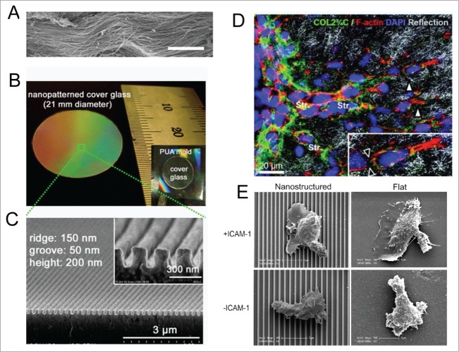
Figure 3.
Cells actively sense and respond to engineered nanotopographical cues. (A) SEM images of ex vivo myocardium of adult rat heart shows aligned collagen fibers running in dense bundles for long distances. Scale bar: 10 μm. (B) Photograph of a large-area anisotropically nanofabricated substratum (ANFS) on a glass coverslip mimicking the collagen fibers in A. (C) Cross-sectional SEM image of the ANFS in B. Reproduced with permission from the National Academy of Sciences. (D) Remodeling of ECM structures by motile HT1080 fibrosarcoma cells. Transition from individual to collective invasion is displayed in 3D spheroids cultured within a 3D collagen lattice. Single cells (white arrowheads) generate small proteolytic tracks (black arrowheads in inset) that become further remodeled and widened by solid strands of multiple cells (Str). The box marks the area magnified in the inset (bottom right); Reprinted with permission from the American Association for Cancer Research. (E) Representative SEM images of T cells on nanostructured (left column) and flat (right column) surfaces in the presence (upper row) and absence (lower row) of ICAM-1; Reprinted with permission from the Journal of Immunology.

