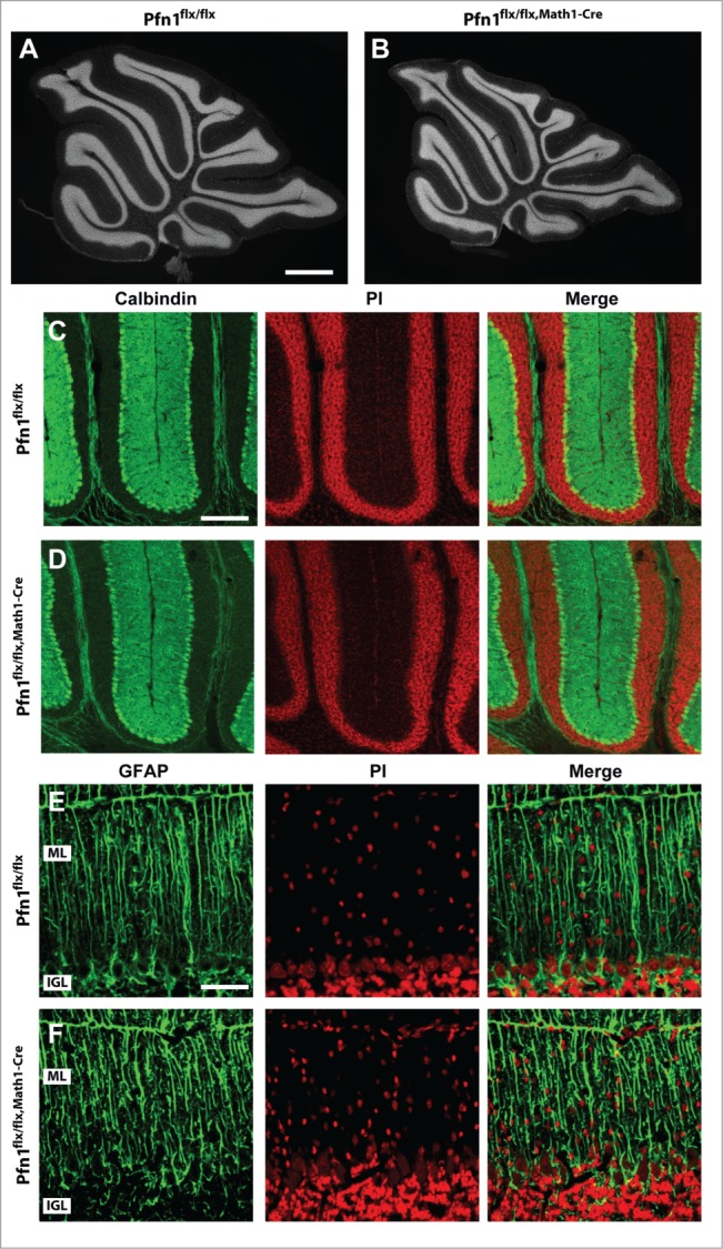
Figure 4.

Normal organization of Purkinje cells and Bergmann glia in Pfn1flx/flx,Math1-Cre mice. (A and B) Propidium iodide-stained cerebellar sections of a Pfn1flx/flx control and a Pfn1flx/flx,Math1-Cre mouse at P30. Scale bar in A corresponds to 1 mm. (C and D) Calbindin immunoreactivity (green) revealed a normal density and organization of Purkinje cells in Pfn1flx/flx,Math1-Cre mice at P30. Sections were counterstained with propidium iodide (PI, red). Scale bar in C corresponds to 100 μm. (E and F) Likewise, the density and organization of Bergmann glia appeared normal in Pfn1flx/flx,Math1-Cre mice, as judged from GFAP immunoreactivity (green). Scale bar in E corresponds to 50 μm.
