
Figure 2.
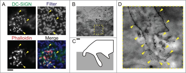
C-type lectins locate to protrusive podosomes-derived structures. (A) Confocal images of human dendritic cells cultured on polycarbonate membrane filters with pore sizes of 1 μm. Actin was stained with phalloidin-Alexa-Fluor-546 (Phalloidin, red), DC-SIGN was labeled by specific primary antibody and secondary antibody conjugated to Alexa-Fluor-488 (DC-SIGN, green). Yellow arrow heads indicate the positioning of protrusion and filter pore. (B–D) Transmission electron microscopy of protrusive podosome-like structures. (B) Electron micrographs of human dendritic cells cultured on polycarbonate filter with 1 μm pore size, impregnated with gelatin and immunogold labeled for CD206. (C) An outline of part of the cell with the protrusion shown in the micrograph in panel (B) (yellow dashed box). (D) Magnification of the protrusion indicated by the yellow box in panel (B). Yellow arrowheads mark positions of clusters of gold-beads which indicate the localization of CD206. Scale bar represents 5 μm (A) and 1 μm (B–D).