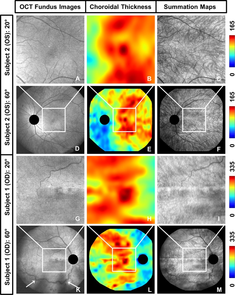
Figure 2.
Additional information provided by wide-field choroidal imaging in comparison to conventional 20° field of view imaging. (A–F) OCT fundus images, ChT maps, and choroidal summation maps showing the left eye of a 34-year-old male diagnosed with retinoschisis (subject 2, spherical equivalent refraction −1.75 D) over a ∼20° field of view (A–C) and a ∼60° field of view (D–F). (G–M) OCT fundus images, ChT maps, and choroidal summation maps showing the right eye of a 63-year-old male with retinal vascular occlusion (subject 1, spherical equivalent refraction +0.5 D) over a ∼20° field of view (G–I) and a ∼60° field of view (K–M). The wide-field images show the detailed ChT and choroidal vascular patterns outside of the macular region and enable examination of the entire region affected by the retinal vascular occlusion in subject 1 (white arrows) (choroidal thickness was measured in μm).

