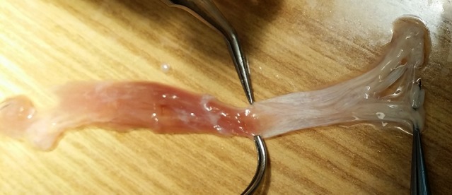
Figure 3.

Isolated human SO muscle and tendon from dissection illustrated in Figure 2, following excision of the trochlea, allowing tendon to unroll flat. Fibers of the medial belly are continuous with the anterior pole of the tendon insertion, while fibers of the lateral muscle belly are continuous with the posterior scleral insertion.
