
Figure 7.
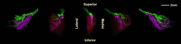
Three-dimensional reconstructions of the superior (green) and inferior (magenta) branches of the intramuscular trochlear nerve in monkey specimen M19. Center image is a posterior view of the nerve with muscle fibers superimposed in red. Flanking images show the nerve and muscle in rotated perspectives 40° and 80° to the left and right.