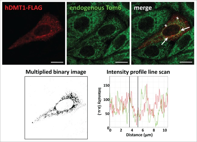
Figure 2.
Transfected hDMT1–1A/+IRE partially colocalizes with endogenous Tom6 in CHO cells. CHO cells were seeded at 4.5 × 10⁴ cells/cm2 on glass coverslips and transiently transfected with hDMT1–1A/+IRE-FLAG 24h later. After 48 hrs of incubation post transfection, they were fixed, permeabilized, blocked, and then stained with primary mouse anti-FLAG (20 μg/ml) (red) and goat anti-Tom6 (1:25) (green) antibodies, followed by secondary Cy3-anti-mouse and Alexa488-anti-goat antibodies. Laser scanning confocal micrographs were acquired sequentially for the different fluorophores. To visualize colocalization, images were merged (top right panel). An intensity profile line scan (bottom right panel) was obtained along the distance indicated by the white crosses in the merged image (top right panel), and a multiplied binary image (bottom left panel) generated as detailed in Materials andMethods. Arrows in the merged image show areas of colocalization of transfected hDMT1–1A/IRE(+) with endogenous Tom-6. Scale bars: 10 μm.

