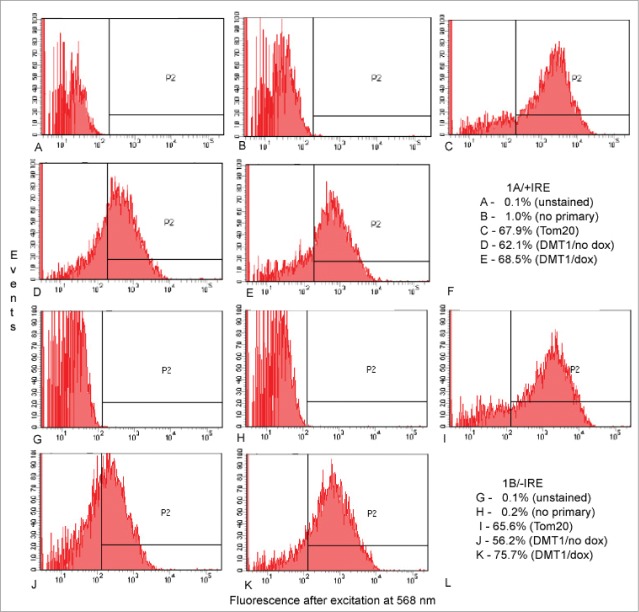
Figure 3.
Detection of DMT1 in isolated mitochondria from HEK293 cells by flow cytometry. Gating on side and forward scatter excluded events smaller than expected for mitochondria as well as those so large that they might represent aggregates of mitochondria or pieces of cell membrane with (A–E) = 1A/+IRE DMT1 and G-K = 1B/-IRE DMT1. Then fluorescence after excitation at 568 nm was determined for (A and G) unstained preparations; (B and H) preparations stained only with secondary antibody; (C and I) preparations stained with anti-Tom20 then secondary antibody; (D and J) preparations from uninduced cells stained with 4EC DMT1 antibody then secondary antibody; (E and K) preparations from doxycycline-induced cells stained with 4EC DMT1 antibody then secondary antibody; (F and L) tabulations of the % of events in the P2 window for (A–E) and (G–K), respectively.

