
Figure 4.
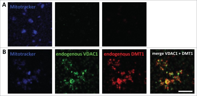
Immunodetection of DMT1 in mitochondria isolated from a rat renal cortical cell line. Mitochondria were isolated from WKPT-0293 Cl.2 rat renal proximal tubule cells as detailed in Materials and Methods. After loading with MitoTracker® Deep Red FM, they were left to attach to coverslips, fixed, permeabilized and blocked. They were then incubated without (A) or with (B) primary goat-anti-VDAC1 and rabbit-anti-DMT1 antibodies, followed by secondary Cy3-anti-rabbit and Alexa488-anti-goat antibodies. Confocal images were acquired and analyzed as described in Materials and Methods. Scale bar: 5 μm.