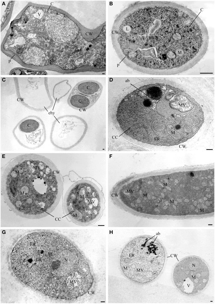
Figure 2.
TEM observations of Sclerotinia sclerotiorum hyphae exposed to bacterial volatiles. (A,B) TEM micrographs showing hyphae ultrastructures of the strain USB-F593 S. sclerotiorum mycelium not exposed to bacterial volatiles. TEM micrographs showing ultrastructural changes of S. sclerotiorum USB-F593 mycelium exposed to volatiles of the Pseudomonas spp. USB2104 (C,D), USB2105 (E,F) and Bacillus spp. USB2103 (G,H). ab, accumulation body; C, cytoplasm; CC, condensed cytoplasm; CW, cell wall; ER, endoplasmic reticulum; ehy, empty hyphae; M, mitochondria; MV, multivesicular; N, nucleus; P, plasmalemma. Bars: 250 nm.

