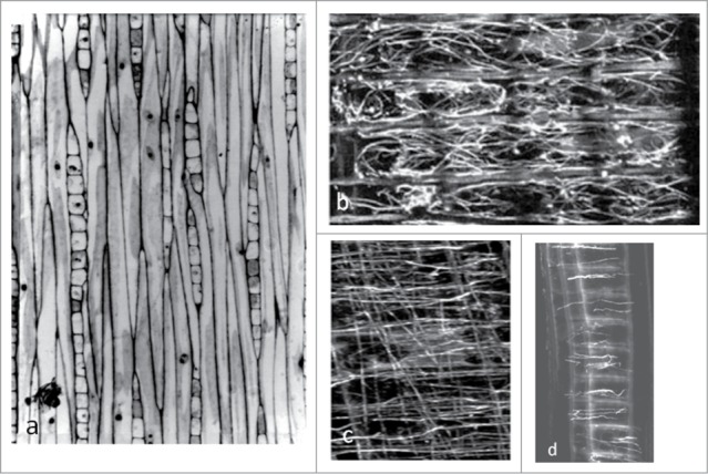
Figure 4.
Ray parenchyma cells within secondary stem tissue of trees. (A) Vertical columns of rays (spindle-shaped groups of small cells) located amongst elongated fusiform cells within the stem cambium of a two-year old hybrid poplar tree. Toluidine blue-stained semi-thin tangential section. Mag. × 280. (B) Radially oriented bundles of MTs in vertical stacks of ray parenchyma cells from Abies sachalinensis. Anti-tubulin immunofluorecent image from a radial section visualised by confocal microscopy. Mag. × 1500. (Micrograph modified from Begum et al.205). (C, D) Radially oriented cables of actin within rays of a two-year old hybrid poplar tree. Anti-actin immunofluorescent image from a radial section visualised by confocal microscopy. Fluorescent images of actin filaments running vertically in c are due to their presence in overlying fusiform cells of the cambium. Mag. × 1200 (C), × 450 (D). (Micrograph modified from Chaffey et al.206).

