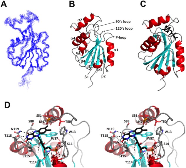
Figure 1.
NMR and crystal structures of Flavodoxin-4 in complex with FMN. (A) Ensemble of 20 energy-minimized CYANA conformers representing the NMR structure (PDB ID 2MWM). (B) Ribbon presentation of the NMR conformer closest to the mean coordinates of the bundle in (A), with identification of the three FMN binding loops. (C) Ribbon presentation of the crystal structure (PDB ID 3EDO). FMN is shown as a black stick diagram. In (B) and (C), the regular secondary structures were defined with DSSP.19(D) Close-up stereo view of the FMN binding site in the crystal structure in (C). Relative to the view in (C) the structure has been rotated by 60° about a vertical axis. Residues in contact with FMN are shown as stick diagrams and identified with the one-letter amino acid code and the sequence number. Dotted black lines indicate hydrogen bonds. The color code for regular secondary structures is used also for the carbon skeleton of the side chains, and oxygen and nitrogen atoms in the side chains and in FMN are colored red and blue, respectively.

