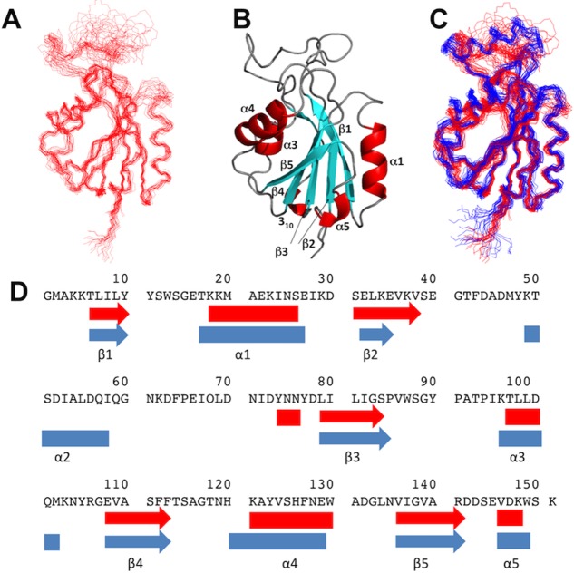
Figure 3.
NMR structure of apo-Flavodoxin-4 and comparison with its FMN complex. (A) Bundle of 20 energy-mimimized CYANA conformers representing the NMR structure. (B) Ribbon presentation of the conformer in (A) which is closest to the mean atom coordinates of the bundle. (C) Superposition of bundles of 20 NMR conformers of apo-Flavodoxin-4 (red) and its FMN-complex (blue). (D) Sequence positions of regular secondary structures in the NMR structures of apo-Flavodoxin-4 (red) and its FMN-complex (blue). Arrows indicate β-strands, and α-helices are shown as rectangles.

