Figure 2.

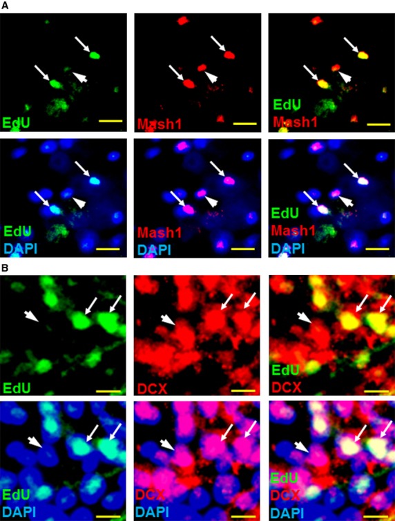
Proliferating spinal cord cells express neurogenic markers. (A) Immunofluorescence analysis showing the overlap of cell proliferation marker EdU (green, FITC) and proneural marker Mash1 (red, Cy3) in spinal cord dorsal horn, week 4 post-CCI, suggests that proliferating adult neural stem cells progress through neuronal differentiation stages similar to embryonic stem cells (arrows). DAPI fluorescence (blue) indicates all nuclei present. Some Mash1+ cells did not stain for EdU, suggesting they had stopped proliferating before EdU treatment (arrowhead). (B) Immunofluorescence analysis showing the overlap of EdU and neurogenesis marker DCX (red, arrows) demonstrate that newly generated NPCs progress through the final stages of neurogenesis within 8 days of EdU treatment. DCX+/EdU- cells (arrowheads) indicate NPCs that had already stopped proliferating before EdU treatment. Scale bars represent 20 μm (A), 10 μm (B).
