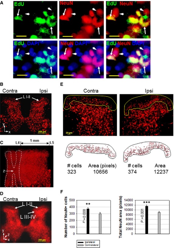
Figure 3.

CCI Generates New Neurons in the Adult Spinal Cord. (A) Co-localization of proliferation marker EdU, neuronal marker NeuN and nuclear stain DAPI in the adult spinal cord (arrows), indicating newly generated neurons 4 weeks post-CCI. Arrowheads indicate cells stained by NeuN only, corresponding to pre-existing neurons. (B) Transversal slice of rat lumbar spinal cord, showing increased NeuN staining in ipsilateral laminae I–II (L I–II, arrows), 6 weeks post-CCI. (C) Stereoscopic reconstruction of a 1 mm-long spinal cord segment (region L4–L5) as a stack of 25 sequential spinal cord slices, rotated 60° around the y-axis (z = longitudinal axis), 6 weeks post-CCI. (D) Transversal view along z axis through the 25-section stack reveals areas with increased NeuN staining density in ipsilateral laminae I–IV (L I–IV) that correspond to areas of nociceptive transmission. (E) Individual slice quantification of ipsilateral/contralateral laminae I–II neurons (yellow perimeter), performed with NIH Image J. The number of NeuN-stained cells and total NeuN-stained areas are shown. (F) Quantification of CCI-induced changes in the number of NeuN-stained cells and in NeuN-stained areas in laminae I–II, 6 weeks post-CCI. Data shown as mean ± SEM, n = 5 rats. Statistically significant differences between ipsilateral versus contralateral laminae I–II (two-tailed t-test) are shown. Scale bars: 10 μm (A), 200 μm (B–D), 20 μm (E).
