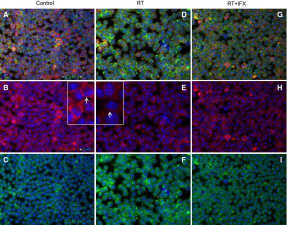
Figure 10.

Double staining of U-937 cells sections with antibodies directed against PECAM-1 (green) and ICAM-1 (red), followed by fluorescence after 4 hrs of treatment. (A–C) control sections (D–F) RT (G–I) RT+IFX. Result show representative picture of four to six slides. Original magnification, ×100.
