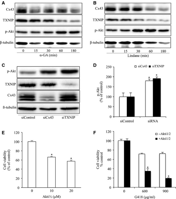
Figure 5.

Inhibition of Cx43 and TXNIP activate AKT. (A and B) Effects of GJ inhibitors on Akt phosphorylation and TXNIP. NRK cells were treated with 7.5 μM α-GA or 100 μM lindane for the indicated time intervals. Cellular lysate were subjected to Western blot analysis of phosphorylated Akt, TXNIP and Cx43. (C and D) Downregulation of Cx43 or TXNIP on Akt phosphorylation. NRK cells were transfected with either Cx43 siRNA or TXNIP siRNA for 48 hrs. Cellular lysates were subjected to Western blot analysis for phosphorylated Akt, TXNIP and Cx43. Equal loading of protein per lane was verified by probing the blots with an anti-β-tubulin antibody. Densitometric analyses of phosphorylated Akt were done by using ImageJ software and are expressed as percentage of the control (mean ± SE, n = 3; *P < 0.05 compared with the siRNA control). (E) Effects of Akt inhibitor on cell viability. NRK cells were incubated with the indicated various concentrations of Akt inhibitor Akti1/2 for 60 hrs. The cell viability was evaluated by CCK-8 assay. Data are expressed as percentage of living cells against the untreated control (mean ± SD, n = 4; *P < 0.05 versus untreated control). (F) Effects of Akt inhibitor on G418-induced cell injury. NRKs were exposed to the indicated concentrations of G418 for 18 hrs in the presence or absence of Akt inhibitor, 10 μM Akti1/2. The cell viability was evaluated by CCK-8 assay. Data are expressed as percentage of living cells against the untreated control (mean ± SD, n = 4; *P < 0.05 versus G418 alone).
