
Fig. 4.
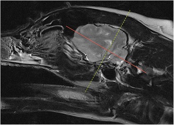
Parasaggital slice in a veterinary epilepsy-specific protocol for 1.5 T MRI scanner. T2W parasagittal image of the brain demonstrating a planned sequence parallel (yellow dotted line) and perpendicular (red solid line) to the long axis of the hippocampus. Images obtained in a 1.5 T MRI (Siemens Symphony, Erlangen, Germany)