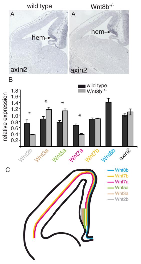
Figure 8. Canonical Wnt signalling appears normal and expression of Wnts 3a and 5a is increased in Wnt8b−/− mutants.
Axin2 in situ hybridisation on coronal sections of telencephalon in E12.5 wild type (A) and Wnt8b−/− mutant embryos (A’). The arrow points to the cortical hem (hem). (B) Quantitative RT-PCR showing the expression levels of Axin2 and several Wnt genes in the E12.5 telencephalon relative to GAPDH. Expression of Wnt3a and Wnt5a is increased in Wnt8b−/− embryos while expression of Wnt2b and Wnt7a is lower. Wnt8b expression is completely lost, as expected. Axin2 levels are unchanged. n=3 embryos of each genotype, * p<0.05, Student’s t-test. (C) Schematic summary of Wnt gene expression in E12.5 dorsal telencephalon. Note that the lines indicate only the extent of expression along the medial-lateral axis – each of these Wnts is expressed throughout the full thickness of the tissue at this stage, except Wnt7b which is expressed throughout the hem but whose cortical expression is restricted to the outermost cells (pial edge) in the cortex.

