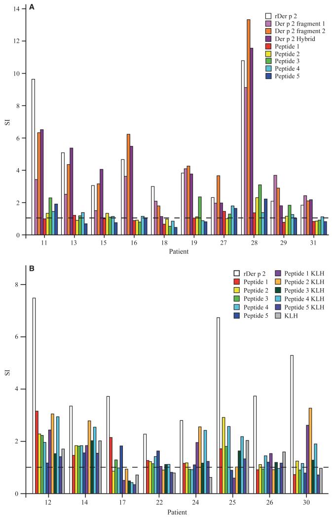
Figure 4.
T-cell proliferative responses of allergic patients to rDer p 2, rDer p 2 derivatives, and Der p 2 peptides. (A) Peripheral blood mononuclear cells (PBMCs) from ten house dust mite (HDM)-allergic patients (x-axis) were stimulated with equimolar amounts of rDer p 2, rDer p 2 derivatives (Der p 2 fragment 1, 2 and hybrid), or Der p 2 peptides. (B) PBMCs from eight additional HDM-allergic patients with different HLA backgrounds (x-axis) were stimulated with equimolar amounts of rDer p 2, unconjugated Der p 2 peptides, keyhole limpet hemocyanin (KLH)-conjugated Der p 2 peptides or KLH. T-cell proliferation was measured by [3H]-thymidine uptake and is displayed as stimulation index (y-axis).

